Ortopædkirurgi

Ortopædkirurgi eller ortopædisk kirurgi (tidligere ortopædi).
Navnet blev konstrueret i 1741 af den franske læge Nicolas Andry ud fra græsk orthos (lige) og paidion (barn), og omhandlede oprindeligt den ikke-kirurgiske behandling af børn med primært skoliose (skæv rygsøjle). Nu omhandler dette lægevidenskabelige speciale den kirurgiske og ikke-kirurgiske behandling af skader samt medfødte og erhvervede lidelser i bevægeapparatet.
Der er i Danmark ni subspecialer:
- Traumatologi – skader inklusive knoglebrud
- Børneortopædi – medfødte misdannelser og visse skader på bevægeapparatet
- Håndkirurgi herunder replantationskirurgi (påsætning afhuggede lemmer)
- Knæ og hoftealloplastik – Indsættelse af et kunstigt led
- Rygkirurgi – Operation af rygskader som knoglebrud, rygdeformiteter som skoliose (skæv rygsøjle) samt rygsygdomme som diskusprolaps
- Tumorkirurgi – Kirurgisk behandling af kræft i bevægeapparatet
- Fod- og ankelkirurgi - Behandling af fod og ankellidelser
- Skulder- og albuekirurgi - Behandling af skader på albue og skulder. Også indsættelse af skulderalloplastik
- Idrætstraumatologi - behandling af idrætsskader som rekonstruktion af forreste korsbånd
I Danmark bemandes den kirurgiske del af skadestuerne med læger fra ortopædkirurgisk afdeling. Ortopædkirurgiens største opgaver er dels behandlingen af knoglebrud (frakturer), dels gipsskinner og ortoser (skinner og korset). Og osteosyntese vha. skruer, skinner, stave m.m. Forud for en ortopædkirurgisk operation foretages ofte en artroskopi. Ortopædiens symbol er et lille skævt træ op ad en stav omviklet med forbinding. Ortopædkirurgien varetages af en speciallæge i ortopædisk kirurgi, en ortopædkirurg.
Se også
 | Wikimedia Commons har medier relateret til: |
Medier brugt på denne side
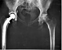
An A-P X-ray of a pelvis showing a total hip joint replacement. The right hip joint (on the left in the photograph) has been replaced. A metal prostheses is cemented in the top of the right femur and the head of the femur has been replaced by the rounded head of the prosthesis. A white plastic cup is cemented into the acetabulum to complete the two surfaces of the artificial "ball and socket" joint.
Although not the case here, hip prostheses can also be made of a ceramic material which rarely wears out during the patient's lifetime. During the operation, a bonding cement is used to fix the metal prostheses into the shaft of the femur and the plastic cup to the acetabulum (socket in the hip bone). One of the leading reasons for hip replacement is osteoarthritis of the hip joint in which virtually all of the cartilage around the top of the femur bone deteriorates due to wear over time, leaving a grinding bone-on-bone situation with the bone surfaces becoming roughened leading to pain and stiffness. Narrowing of joint space (the space between the acetabulum and the head of the femur) is also a feature of osteoarthritis. There may be other changes which are not entirely clear on this A-P X-ray, which probably should be reported together with lateral X-rays of the hips, or with modern computerised imaging techniques.
Keywords: total hip replacement, prosthesis, osteoarthritis, X-ray.