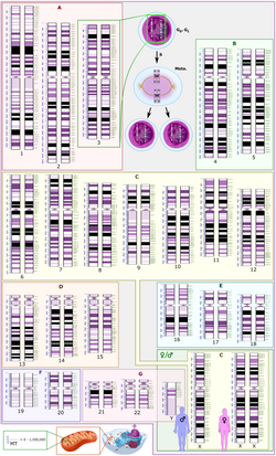
Karyotype
En karyotype er en angivelse af den specifikke kromosombesætning hos eukaryote arter.[1][2] Begrebet kan også anvendes om den enkelte celle eller det enkelte individ.[3] Studiet af karyotyper er en del af cytogenetikken.
Antallet af kromosomer i de somatiske celler hos et individ eller en art kaldes det somatiske antal og betegnes 2n. Således svarer 2n til 46 kromosomer hos mennesket. I kønscellerne er antallet af kromosomer n (hos mennesket er n = 23).[1]
Således er autosomerne i normale, diploide organismer til stede i 2 kopier. Der kan i nogle tilfælde også være kønskromosomer. Polyploide celler har flere kopier af kromosomerne og haploide celler har kun et af hvert kromosom. Studiet af karyotyper er kendt under navnet karyologi. Kromosomerne afbildes normalvis i et karyogram eller idiogram i par ordnet efter størrelse og positionen af centromeret for kromosomer af samme størrelse.
Karyotyper kan anvendes til mange formål såsom at studere fejl i kromosomerne, cellers funktion, taksonomiske forhold, og til at indsamle information om evolutionære begivenheder.
Karyotyper hos mennesket
De fleste arter har en standardkaryotype. Den normale karyotype hos mennesket indeholder 22 par autosomer og et par kønskromosomer. Den normale karyotype hos kvinder indeholder to x-kromosomer og betegnes 46,XX. Mandens karyotype omfatter både et X- og et Y-kromosom og betegnes således 46,XY. Enhver afvigelse fra den normale karyotype kan føre til udviklingsforstyrrelser.
Se også
Referencer
- ^ a b White M.J.D. 1973. The chromosomes. 6th ed, Chapman & Hall, London. p28
- ^ Stebbins G.L. 1950. Variation and evolution in plants. Chapter XII: The Karyotype. Columbia University Press N.Y.
- ^ King R.C., Stansfield W.D. and Mulligan P.K. 2006. A dictionary of genetics. 7th ed, Oxford U.P Oxford & N.Y. p242
Medier brugt på denne side
Forfatter/Opretter:

- Reusing images
- Conflicts of interest:
None
- The work integrates Public Domain source images by Was A Bee, Kelvin Ma and Michał Komorniczak
- Also, a special acknowledgement to everyone involved in the Human Genome Project for generating the source data.
, Licens: CC0
Schematic and relatively language-neutral karyogram of a human. Even at low magnification, it gives an overview of the human genome, with numbered chromosome pairs, its main changes during the cell cycle (top center), and the mitochondrial genome to scale (at bottom left). High magnification (of full size version) more clearly depicts a scale to the left of each chromosome pair that shows the length in terms of million base pairs, and the scale to the right of each chromosome pair shows the designations of the bands and sub-bands. It shows darker and brighter regions as seen on G banding. Each row is vertically aligned at centromere level. The chromosomes are organized into the groups A to G in regard to size, position of the centromere and sometimes the presence of a chromosomal satellite. The chromosome copy numbers are those seen in G0 and G1 of the cell cycle, and at top center it also shows the state of the chromosome 3 pair in the metaphase of the cell cycle, after the synthesis of a copy of each chromosome. The karyogram shows:
- 22 homologous autosomal chromosome pairs
- Both the female (XX) and male (XY) versions of the two sex chromosomes (in green rectangle at bottom right).
- The human mitochondrial genome to scale (at bottom left).
- For more information and context, see Wikipedia:Karyotype.
Karyotype of a human male.


