Hvid substans
| Hvid suubstans | |
|---|---|
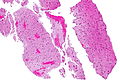
 Micrograf der viser hvid substans med dets karakteristiske fine netagtige udseende (venstre for billedet, lysere pink) og grå substans, med dets karakteristiske neuronale cellekroppe (højre for billedet - mørkere pink). HPS farvning. | |
 Dissekterede menneskelig hjerne, lateralt vue, der viser grå substans (de mørkere dele), og hvid substans (den indre og prominenter hvidere dele). | |
| Detaljer | |
| Identifikatorer | |
| Latin | substantia alba |
| Dorlands /Elsevier | White matter |
| TA | A14.1.00.009 A14.1.02.024 A14.1.02.201 A14.1.04.101 A14.1.05.102 A14.1.05.302 A14.1.06.201 |
| FMA | 83929 |
| Anatomisk terminologi | |
Hvid substans er en af de to komponenter i centralnervesystemet og består mestendels af myelinerede aksoner. Den anden komponent i systemet er grå substans.
| ||||||||||||||||||||||||||||||||||||||||||||||||||||||||||||||||||||||||||||
| Spire Denne artikel om anatomi er en spire som bør udbygges. Du er velkommen til at hjælpe Wikipedia ved at udvide den. |
Medier brugt på denne side
Human brain right dissected lateral view description.JPG
Forfatter/Opretter:
Forfatter/Opretter:
John A Beal, PhD
Dep't. of Cellular Biology & Anatomy, Louisiana State University Health Sciences Center Shreveport, Licens: CC BY 2.5Human brain right dissected lateral view description.JPG
Lateral Portion of Frontal, Parietal, Occipital, and Superior Portion of Temporal Lobe Resected.
The anterior horn of the lateral ventricle is located in the frontal lobe. The body of the lateral ventricle continues posteriorly into the parietal lobe, the posterior horn into the occipital lobe, and the inferior horn down into the temporal lobe. Some structures produce elevations or bumps in the walls of the posterior and/or inferior horns of the lateral ventricles.
- Ventriculus lateralis, Cornu frontale
- Ventriculus lateralis, Pars centralis
- Calcar avis
- Ventriculus lateralis, Cornu occipitale
- Trigonum collaterale
- Eminentia collateralis
- Hippocampus
- Ventriculus lateralis, Cornu temporale
- Capsula interna
- Nucleus caudatus
Grey matter and white matter - very high mag.jpg
Forfatter/Opretter: Nephron, Licens: CC BY-SA 3.0
Very high magnification micrograph of brain, showing normal white matter and normal grey matter. HPS stain. Brain biopsy.
Forfatter/Opretter: Nephron, Licens: CC BY-SA 3.0
Very high magnification micrograph of brain, showing normal white matter and normal grey matter. HPS stain. Brain biopsy.
Related images
-
 Intermed. mag.
 -
 High mag.
 -
 Very high mag.


