Human coronavirus HKU1
| Human coronavirus HKU1 (HCoV-HKU1) | |
|---|---|
 Formation af HCoV-HKU1 | |
| Videnskabelig klassifikation | |
| Domæne | Riboviria |
| Rige | Orthornavirae |
| Række | Pisuviricota |
| Klasse | Pisoniviricetes |
| Orden | Nidovirales |
| Familie | Coronaviridae |
| Slægt | Betacoronavirus |
| Underslægt | Embecovirus |
| Art | Human coronavirus HKU1 |
| Hjælp til læsning af taksobokse | |
Human coronavirus HKU1 (HCoV-HKU1) er en art af coronavirus, der stammer fra inficerede mus.[1] Hos mennesker kan den resultere i infektion i de øvre luftveje med symptomer på forkølelse, men kan udvikle sig til lungebetændelse og bronchiolitis.[1]
Den blev først opdaget i januar 2005 hos to patienter i Hong Kong.[2] Efterfølgende forskning har vist, at den har global udbredelse og en tidligere udviklingshistorie.
Virussen er en indkapslet, enkeltstrenget (single-stranded) RNA-virus med positiv sense, der kommer ind i værtscellen ved at binde til receptoren 'N-acetyl-9-O-acetylneuraminsyre'.[1] Den har genet for Hemagglutinin esterase (HE), et kendetegn for medlemmer af slægten Betacoronavirus og underslægten Embecovirus.[3]
HCoV-HKU1 er en af syv kendte coronavirus der kan inficere mennesker; de øvrige er: HCoV-229E, HCoV-NL63, HCoV-OC43, MERS-CoV, SARS-CoV og SARS-CoV-2.
Referencer
- ^ a b c Lim, Yvonne Xinyi; Ng, Yan Ling; Tam, James P.; Liu, Ding Xiang (2016-07-25). "Human Coronaviruses: A Review of Virus–Host Interactions". Diseases. 4 (3): 26. doi:10.3390/diseases4030026. ISSN 2079-9721. PMC 5456285. PMID 28933406.
Vedrørende mus som vært ('Host: Mice'). Se side 3 i pdf-fil: 'Table 1. Classification of human coronavirus'
{{cite journal}}: Ekstern henvisning i|quote= - ^ Lau, S. K. P.; Woo, P. C. Y.; Yip, C. C. Y.; Tse, H.; Tsoi, H.-w.; Cheng, V. C. C.; Lee, P.; Tang, B. S. F.; Cheung, C. H. Y.; Lee, R. A.; So, L.-y.; Lau, Y.-l.; Chan, K.-h.; Yuen, K.-y. (2006). "Coronavirus HKU1 and Other Coronavirus Infections in Hong Kong". Journal of Clinical Microbiology. 44 (6): 2063-71. doi:10.1128/JCM.02614-05. PMC 1489438. PMID 16757599.
- ^ Woo, Patrick C. Y.; Huang, Yi; Lau, Susanna K. P.; Yuen, Kwok-Yung (2010-08-24). "Coronavirus Genomics and Bioinformatics Analysis". Viruses. 2 (8): 1804-1820. doi:10.3390/v2081803. ISSN 1999-4915. PMC 3185738. PMID 21994708.
In all members of Betacoronavirus subgroup A, a haemagglutinin esterase (HE) gene, which encodes a glycoprotein with neuraminate O-acetyl-esterase activity and the active site FGDS, is present downstream to ORF1ab and upstream to S gene (Figure 1).
Se også
Eksterne henvisninger
 Wikispecies har taksonomi med forbindelse til Human coronavirus HKU1
- "Coronaviruses" fra Micro.msb.le.ac.uk
- "Betacoronavirus" fra Viralzone.expasy.org
- 'Taxon identifiers'. Engelsk hjælpeside til 'taksonindentifikatorer'
| Spire Denne artikel om biologi er en spire som bør udbygges. Du er velkommen til at hjælpe Wikipedia ved at udvide den. |
Medier brugt på denne side
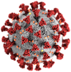
This illustration, created at the Centers for Disease Control and Prevention (CDC), reveals ultrastructural morphology exhibited by coronaviruses. Note the spikes that adorn the outer surface of the virus, which impart the look of a corona surrounding the virion, when viewed electron microscopically. A novel coronavirus, named Severe Acute Respiratory Syndrome coronavirus 2 (SARS-CoV-2), was identified as the cause of an outbreak of respiratory illness first detected in Wuhan, China in 2019. The illness caused by this virus has been named coronavirus disease 2019 (COVID-19).
Forfatter/Opretter: Emmanuel Boutet, Licens: CC BY-SA 2.5
A black bee (Apis mellifera mellifera.
Forfatter/Opretter: © 2013 Dominguez et al., Licens: CC BY 4.0
Formation of large syncytia of primary human alveolar type II cells infected with HCoV-HKU1. Cells were inoculated with a 1∶10 dilution of the clinical isolate HKU1/DEN/2010/21 at 34oC and fixed 120 hours post infection. Type II cell cultures were immunolabeled with polyclonal rabbit antibodies to purified HCoV-HKU1 spike protein and fluorescein labeled anti rabbit IgG (green). Nuclei were stained with DAPI (blue). Viral antigen is seen only within the cytoplasm of the cells. Efficient infection with cell to cell spread and formation of large, multinucleated giant cells is clearly evident.


