Granulocyt


Granulocytter er en undergruppe af de hvide blodlegemer (leukocytter), og hører sammen med røde blodlegemer (erytrocytter) og blodplader (trombocytter) til blodets formede elementer.
Der findes 5 forskellige slags leukocytter i blodet, hvoraf de granulære leukocytter udgør de tre, og lymfocytten og monocytten de to øvrige. Granulocytterne adskilles fra de øvrige ved deres granulære cytoplasmatiske udseende i et lysmikroskop.
Granulocytterne inddeles yderligere efter deres farvbarhed i deres cytoplasmatiske granula:
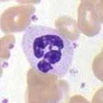
Neutrofile granulocytter
Struktur: Neutrofile granulocytter ses som en 10-15 mikrometer stor celle, med en kerne med 3-5 lapper. Cytoplasmaet indeholder talrige granula. Disse indeholder bl.a. myeloperoxid, sure hydrolaser og lysozym.
Funktion: De neutrofile granulocytter er sammen med makrofagerne kroppens primære fagocytterende celler. Deres funktion er primært at fagocyttere bakterier, og derved bekæmpe infektioner.

Basofile granulocytter
Struktur: De basofile granulocytter er ca. 12-15 mikrometer store celler, med en 2-3 lappet kerne, som evt. er S-formet. Indeholder i sit cytoplasma store basofile granula, der bl.a. indeholder heparin, histamin, lysosomale enzymer samt peroxidase.
Funktion: Det vides ikke med sikkerhed hvad de basofiles præcise funktion er i kredsløbet. Det vides dog at de har store lighedspunkter med mastcellerne. Muligvis er de en slags forstadie til disse, som så forlader kredsløbet og deltager i anafylaktiske reaktioner.
Eosinofile granulocytter

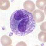
Struktur: Er ligesom de andre granulocytter 12-15 mikrometer i diameter. Den eosinofiles kerne ses som 2 store lapper. Indeholder membranafgrænsede granula der indeholder meoloperoxidase og lysosomale enzymer. Cytoplasmaets granula er kraftigt eosint.
Funktion: Deres vigtigste funktion er bekæmpelse af parasitære infektioner.
Se også
Medier brugt på denne side
Neutrophile segmented Granulocyte
Basophile Granulocyte
Forfatter/Opretter:

- Reusing images
- Conflicts of interest:
None
This diagram shows the hematopoiesis as it occurs in humans. It may look incomplete when rendered directly from WikiMedia. Reference list is found at: File:Hematopoiesis (human) diagram.png
- The morphological characteristics of the hematopoietic cells are shown as seen in a Wright’s stain, May-Giemsa stain or May-Grünwald-Giemsa stain. Alternative names of certain cells are indicated between parentheses.
- Certain cells may have more than one characteristic appearance. In these cases, more than one representation of the same cell has been included.
- Together, the monocyte and the lymphocytes comprise the agranulocytes, as opposed to the granulocytes (basophil, neutrophil and eosinophil) that are produced during granulopoiesis.
- B., N. and E. stand for Basophilic, Neutrophilic and Eosinophilic, respectively – as in Basophilic promyelocyte. For lymphocytes, the T and B are actual designations.
[1] The polychromatic erythrocyte (reticulocyte) at the right shows its characteristic appearance when stained with methylene blue or Azure B.
[2] The erythrocyte at the right is a more accurate representation of its appearance in reality when viewed through a microscope.
[3] Other cells that arise from the monocyte: osteoclast, microglia (central nervous system), Langerhans cell (epidermis), Kupffer cell (liver).
Eosinophile Granulocyte


