Ebolavirus


Ebolavirus, EBOV, er en virus, der forårsager Ebola (en. Ebola virus disease (EVD) eller tidligere Ebola haemorrhagic fever (EHF)), en zoonose, der spredes fra flagermus til mennesker.
Ebolavirus kaldes sammen med Marburgvirus og Lassavirus for de afrikanske blødningsfebervirus og klassificeres som virus i højeste fareklasse.[1][2]
EBOV er en trådformet, membrankappet virus med et genom af -ssRNA og er en del af Filoviridae-familien, nært beslægtet med Marburgvirus.
Historisk
EBOV er opkaldt efter floden Ebola i DR Congo, hvor virussen også blev opdaget første gang. Fra 1976 har der været gentagne udbrud af Ebola. To stammer blev identificeret, hhv. fra Zaire og fra Sudan med gennemsnitlige dødelighedsprocenter på henholdsvis 83% og 54%. En tredje stamme, Reston-Ebolavirus, blev opdaget i 1989 i en gruppe af aber (Macaca fascicularis) importeret fra Filippinerne til Hazleton Primate Quarantine Unit i Reston, Virginia i USA. Denne stamme har ikke været humanpatogen. Der kendes nu fem stammer af Ebolavirus (BDBV, EBOV, RESTV, SUDV and TAFV).
Generelt
I elektronmikroskop fremtræder EBOV som langstrakte, fleksible tråde med en diameter på 80 nm og en længde fra 800 til 1400 nm.
Genom
EBOV’s genom er -ssRNA (negativt polariseret enkeltstrænget RNA) på 18-19.000 nukleotider, der koder for 7 proteiner.[3][4] De fem forskellige stammer afviger i nukleotidsekvensen og antallet og placeringen af overlappende gener.
Proteiner
- Glycoprotein, multifunktionelt overfladeprotein GP, sGP, ssGP, GP1 og GP2
- Matrixprotein VP40, hexamer
- RNA dependent RNA polymerase, L protein
- Nucleoprotein NP
- Polymerase cofactor VP35 (multifunktionelt nucleocapsidprotein)
- Transcription activator VP30 (nucleocapsidprotein)
Receptorer for EBOV
EBOV glycoprotein GP binder sig til
- TIM-1, et overfladeprotein på værtsorganismens epithelceller [9]
- C-type lectinerne DC-SIGN, L-SIGN og hMGL på overfladen af værtsorganismens monocyter, dendritceller og makrofager
- Niemann–Pick C1 (NPC1), et 13TM membranprotein i værtsorganismens endosomer [10]
Vaccine
En Ebolavaccine blev godkendt i USA i 2019.
Se også
Henvisninger
- ^ "Ebola. Statens Seruminstitut". Arkiveret fra originalen 22. juni 2020. Hentet 2. juni 2020.
- ^ "Derfor har Ebola-virus ikke spredt sig. Videnskab.dk". Arkiveret fra originalen 26. juni 2020. Hentet 26. juni 2020.
- ^ "Ebolavirus. Viral Zone". Arkiveret fra originalen 16. marts 2020. Hentet 5. maj 2020.
- ^ "Viruses ARE alive, and they're older than modern cells, new study suggests. Science Alert 2015". Arkiveret fra originalen 28. september 2015. Hentet 5. maj 2020.
- ^ Re-Emerging Human Viral Hemorrhagic Fevers: A Review. American Journal of Infectious Diseases and Microbiology 2016
- ^ "Ebola Virus's Glycoproteins and Entry Mechanism. 2016". Arkiveret fra originalen 8. juni 2020. Hentet 5. maj 2020.
- ^ "Ebola Virus Proteins. Molecule of the Month". Arkiveret fra originalen 30. april 2020. Hentet 5. maj 2020.
- ^ "Structure of Ebola Virus. Microbiology Info 2018". Arkiveret fra originalen 29. september 2020. Hentet 5. maj 2020.
- ^ "Receptor for Ebola virus identified. Science Daily 2011". Arkiveret fra originalen 6. september 2015. Hentet 5. maj 2020.
- ^ "Small molecule inhibitors reveal Niemann-Pick C1 is essential for ebolavirus infection. Nature 2011". Arkiveret fra originalen 12. november 2020. Hentet 5. maj 2020.
|
Medier brugt på denne side
An electron micrograph of an Ebola viral particle showing the characteristic filamentous structure of a Filoviridae. The viral filaments can appear in images in various shapes including a 'u', '6', a coil, or branched resulting in pleomorphic particles. The filaments are reported to be between 60-80 nm in diameter, the length of a filament associated with an individual viral partial is extremely variable with Ebola particles of up to 14,000 nm in length being reported. An average length, which may represent the most infectious particles is in the region of 1000 nm.
The first electronmicrograph of Ebola was 13 October 1976 by Dr. F.A. Murphy, now at UC Davis, who was then working at the CDC.
The nucleocapsid structure consists of a central channel, 20-30nm in diameter, surrounded by helically wound capsid with a diameter of 40-50nm and an interval of 5nm. 7nm glycoprotein spikes spaced 10 nm apart from each other are present within the outer envelope of the virus which is derived from the host cell membrane. Each viral particle contains one molecule of single-stranded, negative-sense RNA, which encodes the seven viral proteins.Ebola virus virion. Created by GC microbiologist Cynthia Goldsmith, this colorized transmission electron micrograph (TEM) revealed some of the ultrastructural morphology displayed by an Ebola virus virion.
Life cycles of the Ebolavirus

Forfatter/Opretter: BernbaumJG, Licens: CC BY 4.0
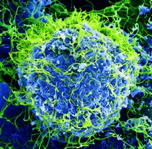
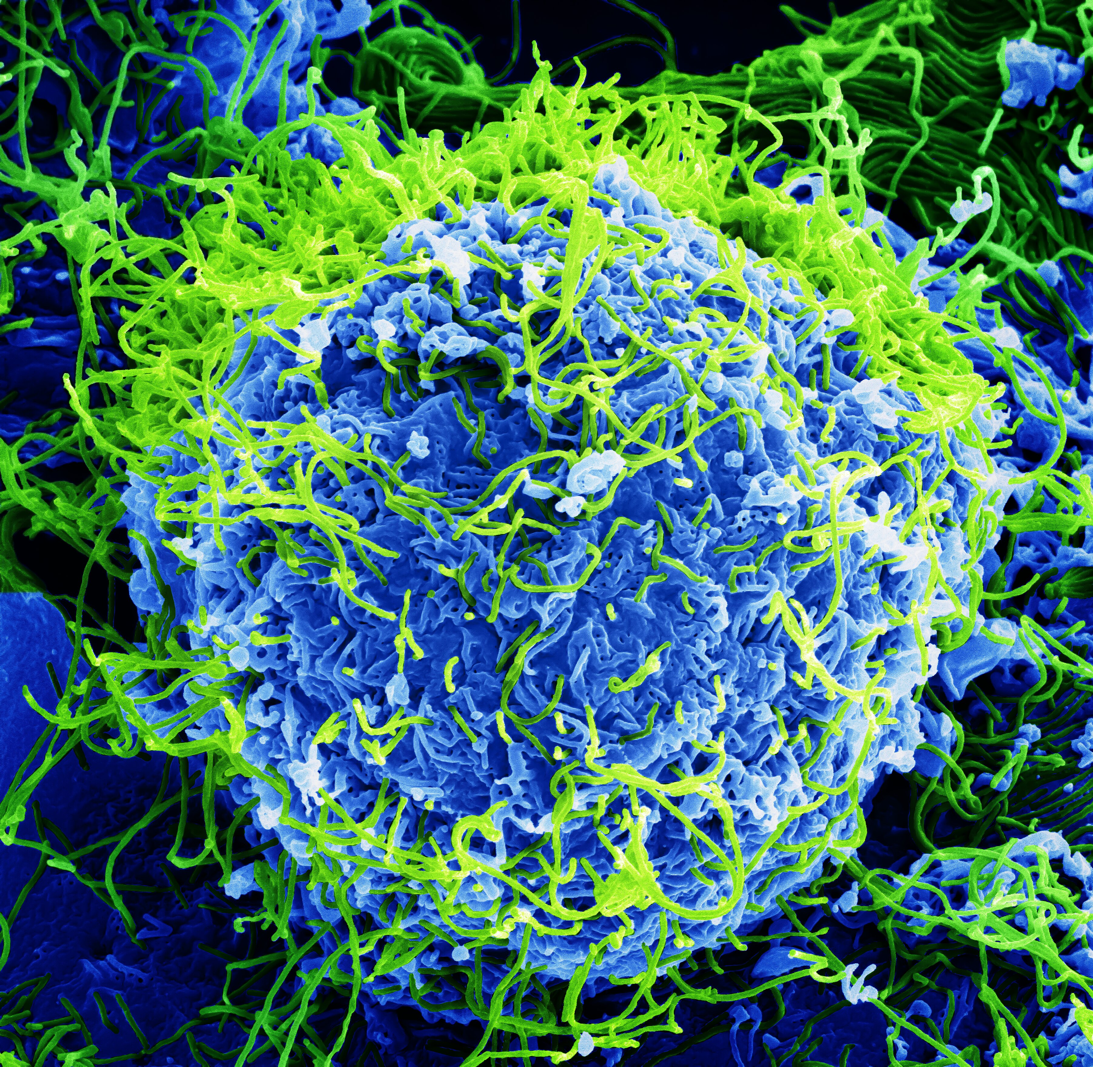
"Beautiful....But Deadly": Colorized scanning electron micrograph of Ebola virus particles (green) found both as extracellular particles and budding particles from a chronically-infected African Green Monkey Kidney cell (blue); 20,000x magnification. JPEG version.