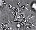
Dendritcelle


Dendritceller (DC) eller antigenpræsenterende celler (APC) er specialiserede leukocytter (hvide blodlegemer), som er en del af pattedyrenes immunforsvar. De findes i huden og de indre overflader af næsen, lungerne, maven og tarmene. Der virker dendritcellerne som mellemled mellem det uspecifikke immunforsvar og det specifikke immunforsvar ved at erkende og optage antigener for derefter at præsentere dem på overfladen over for B-celler og T-celler i en lymfeknudene.[1]
Dendritceller udvikles fra monocytter, der udvikles fra stamceller i benmarven.
Ralph M. Steinman fik i 2011 halvdelen af Nobelprisen i fysiologi eller medicin for at opdage den centrale rolle af dendritcellerne i det uspecifikke immunforsvar.[2]
Mekanisme
Umodne dendritceller reagerer ved hjælp af såkaldte pattern recognition receptorer (PRR) såsom de Toll-lignende receptorer (TLR) på celleoverfladen og på de indre membraner med indtrængende bakterier og virus. Ved pinocytose bliver antigenerne optaget og nedbrudt for at blive præsenteret som fragmenter på MHC-molekylerne på celleoverfladen. Samtidig aktiveres celleoverflade-receptorer kaldet immun-checkpoints, der aktiverer T-cellerne og B-cellerne.
Typer
Konventionelle dendritceller (cDC, tidligere kaldet myeloide dendritceller, mDC) har Toll-lignende receptorer på overfladen (TLR2 og TLR4) og producerer interleukiner, specielt IL6 og IL12, tumor necrosis faktor (TNF) og chemokiner.
Plasmacytoide dendritceller (pDC) har Toll-lignende receptorer på indre membraner (TLR7 og TLR9) og producerer Interferon-alfa (INFa eller type1-Interferon). TLR 7 and TLR 9 reagerer henholdsvis med enkeltstrenget RNA (ssRNA) og ikke-methylerede CpG DNA-sekvencer.
Se også
Henvisninger
- ^ Dendritic Cells Revisited. Annual Review of Immunology 2021
- ^ "Ralph M. Steinman, Facts. The Nobel Prize 2011". Arkiveret fra originalen 14. december 2018. Hentet 14. december 2018.
Medier brugt på denne side
Forfatter/Opretter: Judith Behnsen, Priyanka Narang, Mike Hasenberg, Frank Gunzer, Ursula Bilitewski, Nina Klippel, Manfred Rohde, Matthias Brock, Axel A. Brakhage, Matthias Gunzer, Licens: CC BY 2.5
A screen clip from a video included in the journal article “Environmental Dimensionality Controls the Interaction of Phagocytes with the Pathogenic Fungi Aspergillus fumigatus and Candida albicans”
A well resolved dendritic cell drags a conidium through a distance of up to 9 μm. The conidium, however, is not phagocytosed by the cell.Section of skin showing large numbers of dendritic (Langerhans) cells in the epidermis. Mycobacterium ulcerans infection, S100 immunoperoxidase stain.
Forfatter/Opretter:

- Reusing images
- Conflicts of interest:
None
This diagram shows the hematopoiesis as it occurs in humans. It may look incomplete when rendered directly from WikiMedia. Reference list is found at: File:Hematopoiesis (human) diagram.png
- The morphological characteristics of the hematopoietic cells are shown as seen in a Wright’s stain, May-Giemsa stain or May-Grünwald-Giemsa stain. Alternative names of certain cells are indicated between parentheses.
- Certain cells may have more than one characteristic appearance. In these cases, more than one representation of the same cell has been included.
- Together, the monocyte and the lymphocytes comprise the agranulocytes, as opposed to the granulocytes (basophil, neutrophil and eosinophil) that are produced during granulopoiesis.
- B., N. and E. stand for Basophilic, Neutrophilic and Eosinophilic, respectively – as in Basophilic promyelocyte. For lymphocytes, the T and B are actual designations.
[1] The polychromatic erythrocyte (reticulocyte) at the right shows its characteristic appearance when stained with methylene blue or Azure B.
[2] The erythrocyte at the right is a more accurate representation of its appearance in reality when viewed through a microscope.
[3] Other cells that arise from the monocyte: osteoclast, microglia (central nervous system), Langerhans cell (epidermis), Kupffer cell (liver).
Title: Dendritic cell revealed
Description: Artistic rendering of the surface of a human dendritic cell illustrating sheet-like processes that fold back onto the membrane surface. When exposed to HIV, these sheets entrap viruses in the vicinity and focus them to contact zones with T-cells targeted for infection. These studies were carried out using ion abrasion scanning electron microscopy, a new technology we have been developing and applying for 3D cellular imaging. Categories: Research in NIH Labs and Clinics Type: Color, Diagram Source: National Cancer Institute (NCI) Creator: Don Bliss, Sriram Subramaniam Date Created: Unknown Date Added: 5/24/2012
Reuse Restrictions: None - This image is in the public domain and can be freely reused. Please credit the source and/or author listed above.


