Human brain right dissected lateral view description
Forfatter/Opretter:
John A Beal, PhD
Dep't. of Cellular Biology & Anatomy, Louisiana State University Health Sciences Center Shreveport
Attribution:
Billedet er tagget "Attribution Required", men der blev ikke angivet nogen tilskrivningsoplysninger. Attributionsparameteren blev sandsynligvis udeladt ved brug af MediaWiki-skabelonen til CC-BY-licenserne. Forfattere og ophavsmænd finder et eksempel på korrekt brug af eksempel her. her.
Kredit:
Shortlink:
kilde:
størrelse:
653 x 413 Pixel (40769 Bytes)
beskrivelse:
Human brain right dissected lateral view description.JPG
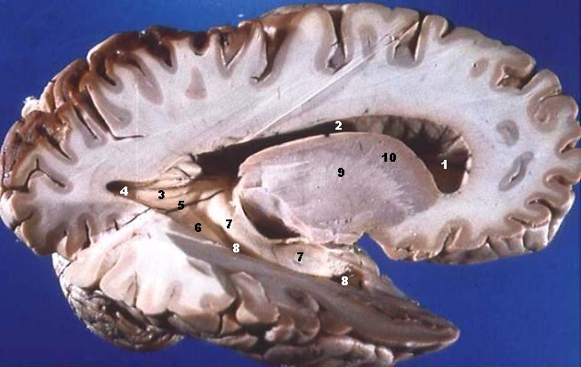
Lateral Portion of Frontal, Parietal, Occipital, and Superior Portion of Temporal Lobe Resected.
The anterior horn of the lateral ventricle is located in the frontal lobe. The body of the lateral ventricle continues posteriorly into the parietal lobe, the posterior horn into the occipital lobe, and the inferior horn down into the temporal lobe. Some structures produce elevations or bumps in the walls of the posterior and/or inferior horns of the lateral ventricles.
- Ventriculus lateralis, Cornu frontale
- Ventriculus lateralis, Pars centralis
- Calcar avis
- Ventriculus lateralis, Cornu occipitale
- Trigonum collaterale
- Eminentia collateralis
- Hippocampus
- Ventriculus lateralis, Cornu temporale
- Capsula interna
- Nucleus caudatus
Licens:
Licensbetingelser:
Creative Commons Attribution 2.5
Yderligere oplysninger om licens til billedet kan findes her. Sidste ændring: Mon, 22 Aug 2022 12:40:32 GMT